Clinical History
A 32-year-old woman presents with a 1.8 cm right thyroid nodule. On ultrasound, the nodule is solid, hypoechoic, and demonstrates microcalcifications. Fine-needle aspiration is performed under ultrasound guidance.
Cytologic Features
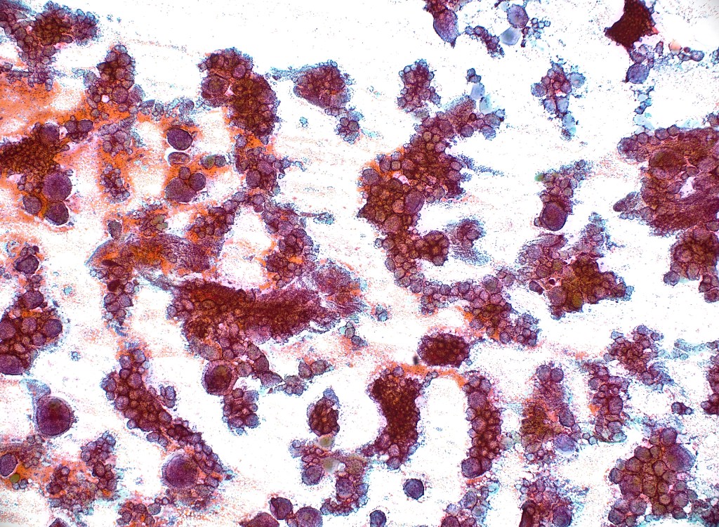
- Moderately cellular specimen
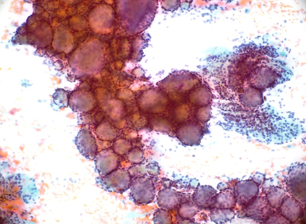
- Papillary tissue fragments
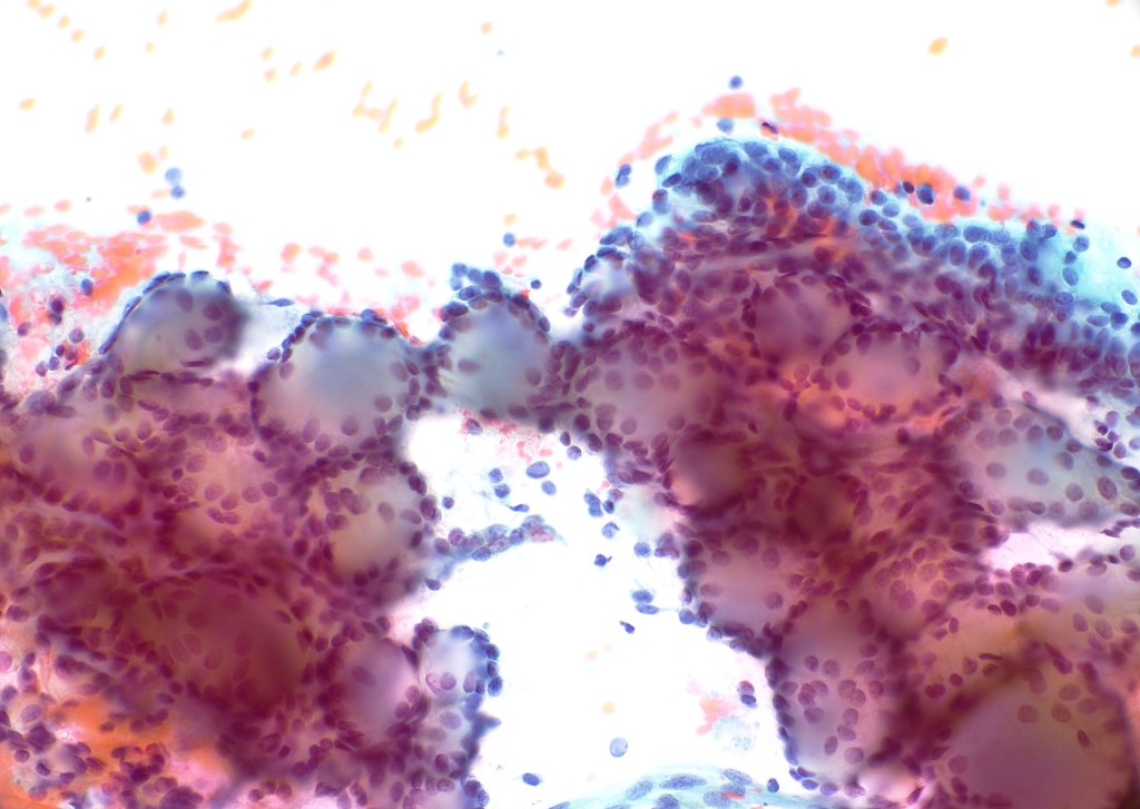
- Enlarged nuclei with overlapping and crowding
- Irregular nuclear contours (important clue)
- Nuclear grooves
- Intranuclear pseudoinclusions
- Pale (optically clear) chromatin
Interpretation
Features consistent with papillary thyroid carcinoma (Bethesda Category VI – Malignant)
Making the Diagnosis
The diagnosis is supported by a moderately cellular aspirate showing true papillary fragments lined by follicular cells demonstrating enlarged, elongated nuclei with irregular membranes, pale chromatin, and nuclear grooves. Occasional intranuclear pseudoinclusions are seen. The chromatin is pale. The nuclear features and architectural pattern are diagnostic of papillary thyroid carcinoma (PTC).
According to the Bethesda System for Reporting Thyroid Cytopathology, this case falls under Category VI, malignant.