Clinical history
A 55-year-old man presents with a slowly enlarging, painless mass in the left submandibular region, present for approximately 8 months. Imaging reveals an ill-defined, solid mass in the submandibular gland. Fine-needle aspiration (FNA) is performed.
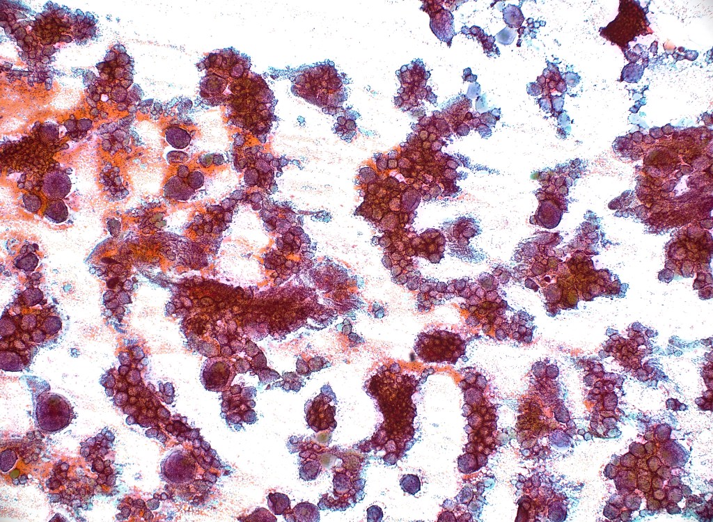
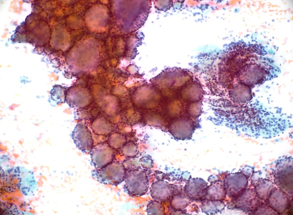
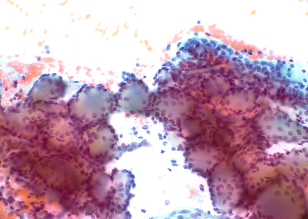
Cytologic Features
- Cellular aspirate
- Small, uniform basaloid cells
- Scant cytoplasm, round to oval nuclei
- Sheets and cribriform arrangements
- Hyaline globules (basement membrane material) within and around cell groups
- No significant pleomorphism
Interpretation
Features consistent with adenoid cystic carcinoma (AdCC)
Making the Diagnosis
The diagnosis is supported by a cellular basaloid neoplasm arranged in predominantly cribriform groups associated with well-defined, rounded hyaline globules present within the clusters. The nuclei are uniform, and the background is clean. These findings are characteristic of adenoid cystic carcinoma. According to the Milan System for Reporting Salivary Gland Cytopathology, this case falls under Category VI – Malignant.
In limited sampling, the differential diagnosis may include basal cell adenoma and other basaloid salivary neoplasms with overlapping features, though the overall pattern here is typical for AdCC.

Leave a comment